- HOME
- News & Events
- [June 24] D5 Medical & Life Science Seminar-Dr. Masahiro Fukuda
News & Events
[June 24] D5 Medical & Life Science Seminar-Dr. Masahiro Fukuda
May 29 2020
The "D5 Medical & Life Science Seminar" course will be offered by International Research Center for Medical Sciences (IRCMS). It will run from April 2020 to March 2021, with lectures given by scientists who are affiliated with IRCMS or in collaboration with researchers at IRCMS. The lectures will be given once a month, in English, and by leading scientists in the relevant research field. Students will be taught: 1) how normal physiological functions are maintained in the human body; 2) how these systems become abnormal under certain pathophysiologic conditions; 3) why stem cells are important in animal development and homeostasis; 4) how stem cell-based approaches can help us understand disease mechanisms and find potential cure for diseases related to stem cell malfunction (e.g., cancer, aging).
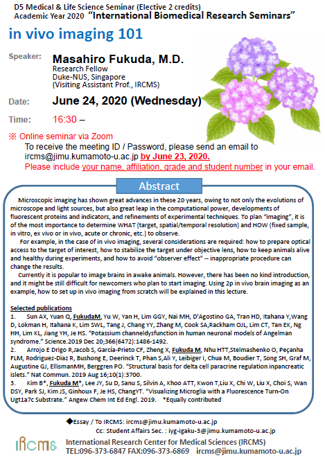
Date : June 24, 2020 (Wednesday)
Time : 16:30 -
* Online seminar via Zoom
To receive the meeting ID / Password, please send an email to ircms@jimu.kumamoto-u.ac.jp
by June 23, 2020. Please include your name, affiliation, grade and student number in your email.
Speaker : Masahiro Fukuda, M.D.
Research Fellow, Duke-NUS, Singapore
Visiting Assistant Prof., IRCMS, Kumamoto University
Title : in vivo imaging 101
Abstract :
Microscopic imaging has shown great advances in these 20 years, owing to not only the evolutions of microscope and light sources, but also great leap in the computational power, developments of fluorescent proteins and indicators, and refinements of experimental techniques. To plan "imaging", it is of the most importance to determine WHAT (target, spatial/temporal resolution) and HOW (fixed sample, in vitro, ex vivo or in vivo, acute or chronic, etc.) to observe.
For example, in the case of in vivo imaging, several considerations are required: how to prepare optical access to the target of interest, how to stabilize the target under objective lens, how to keep animals alive and healthy during experiments, and how to avoid "observer effect" -- inappropriate procedure can change the results.
Currently it is popular to image brains in awake animals. However, there has been no kind introduction, and it might be still difficult for newcomers who plan to start imaging. Using 2p in vivo brain imaging as an example, how to set up in vivo imaging from scratch will be explained in this lecture.
Selected publications
- Sun AX, Yuan Q, Fukuda M, Yu W, Yan H, Lim GGY, Nai MH, D'Agostino GA, Tran HD, Itahana Y,Wang D, Lokman H, Itahana K, Lim SWL, Tang J, Chang YY, Zhang M, Cook SA,Rackham OJL, Lim CT, Tan EK, Ng HH, Lim KL, Jiang YH, Je HS. "Potassium channeldysfunction in human neuronal models of Angelman syndrome." Science.2019 Dec 20;366(6472):1486-1492.
- Arrojo E Drigo R,Jacob S, García-Prieto CF, Zheng X, Fukuda M, Nhu HTT,Stelmashenko O, Peçanha FLM, Rodriguez-Diaz R, Bushong E, Deerinck T, Phan S,Ali Y, Leibiger I, Chua M, Boudier T, Song SH, Graf M, Augustine GJ, EllismanMH, Berggren PO. "Structural basis for delta cell paracrine regulation inpancreatic islets." Nat Commun. 2019 Aug 16;10(1):3700.
- Kim B*, Fukuda M*, Lee JY, Su D, Sanu S, Silvin A, Khoo ATT, Kwon T,Liu X, Chi W, Liu X, Choi S, Wan DSY, Park SJ, Kim JS, Ginhoux F, Je HS, ChangYT. "Visualizing Microglia with a Fluorescence Turn-On Ugt1a7c Substrate." Angew Chem Int Ed Engl. 2019. *Equally contribute

