
- HOME
- News & Events
- [October 9] D5 Medical & Life Science Seminar - Dr. Pilhan Kim
News & Events
[October 9] D5 Medical & Life Science Seminar - Dr. Pilhan Kim
August 28 2019
The "D5 Medical & Life Science Seminar" course will be offered by International Research Center for Medical Sciences (IRCMS). It will run from April 2019 to March 2020, with lectures given by scientists who are affiliated with IRCMS or in collaboration with researchers at IRCMS. The lectures will be given once a month, in English, and by leading scientists in the relevant research field. Students will be taught: 1) how normal physiological functions are maintained in the human body; 2) how abnormalities in these systems (e.g., cancer) are studied using experimental models; 3) cutting-edge technologies (including single cell level imaging and omics analysis) used for mechanistic understanding of these abnormalities; 4) efforts and progresses in finding cure for human diseases associated with these abnormalities; and 5) importance of understanding disease mechanisms using cross-disciplinary approaches.
Date : October 9, 2019 (Wednesday)
Time : 17:30 -
Venue : IRCMS 1F Meeting Lounge
Speaker : Pilhan Kim, Ph.D.
Associate Professor
Graduate School of Medical Science and Engineering (GSMSE)
Graduate School of Nanoscience and Technology (GSNT)
Korea Advanced Institute of Science and Technology (KAIST)
Visiting Associate Professor (IRCMS)
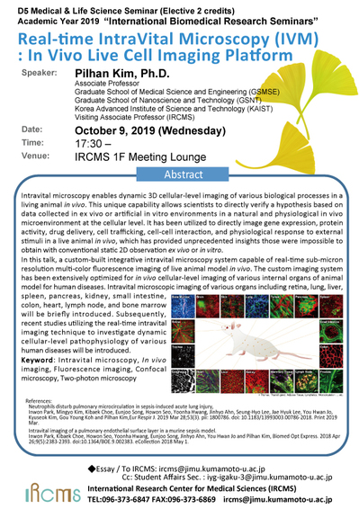
Title : Real-time IntraVital Microscopy (IVM) : In Vivo Live Cell Imaging Platform
Abstract :
Intravital microscopy enables dynamic 3D cellular-level imaging of various biological processes in a living animal in vivo. This unique capability allows scientists to directly verify a hypothesis based on data collected in ex vivo or artificial in vitro environments in a natural and physiological in vivo microenvironment at the cellular level. It has been utilized to directly image gene expression, protein activity, drug delivery, cell trafficking, cell-cell interaction, and physiological response to external stimuli in a live animal in vivo, which has provided unprecedented insights those were impossible to obtain with conventional static 2D observation ex vivo or in vitro.
In this talk, a custom-built integrative intravital microscopy system capable of real-time sub-micron resolution multi-color fluorescence imaging of live animal model in vivo. The custom imaging system has been extensively optimized for in vivo cellular-level imaging of various internal organs of animal model for human diseases. Intravital microscopic imaging of various organs including retina, lung, liver, spleen, pancreas, kidney, small intestine, colon, heart, lymph node, and bone marrow will be briefly introduced. Subsequently, recent studies utilizing the real-time intravital imaging technique to investigate dynamic cellular-level pathophysiology of various human diseases will be introduced.
Keyword: Intravital microscopy, In vivo imaging, Fluorescence imaging, Confocal microscopy, Two-photon microscopy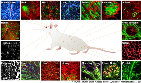
![]()