- HOME
- News & Events
- [Nov 22]*D5 Medical & Life Science Seminar*
News & Events
[Nov 22]*D5 Medical & Life Science Seminar*
November 22 2017
Date: November 22, 2017 (Wed)
Time: 17:30 -
Venue: IRCMS 1F Meeting Lounge
Speaker: Pilhan Kim, Ph.D.
Graduate School of Nanoscience and Technology (GSNT),
Korea Advanced Institute of Science and Technology(KAIST)
(Visiting Associate Professor, IRCMS, Kumamoto University, Japan )
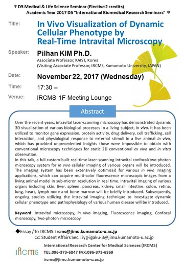
In Vivo Visualization of Dynamic Cellular Phenotype by Real-Time Intravital Microscopy Platform
Over the recent years, intravital laser-scanning microscopy has demonstrated dynamic 3D visualization of various biological processes in a living subject, in vivo. It has been utilized to monitor gene expression, protein activity, drug delivery, cell trafficking, cell interaction, and physiological response to external stimuli in a live animal in vivo, which has provided unprecedented insights those were impossible to obtain with conventional microscopy techniques for static 2D conventional ex vivo and in vitro observation.
In this talk, a full custom-built real-time laser-scanning intravital confocal/two-photon microscopy system for in vivo cellular imaging of various organs will be introduced. The imaging system has been extensively optimized for various in vivo imaging applications, which can acquire multi-color fluorescence microscopic images from a living animal model in sub-micron resolution in real time. Intravital imaging of various organs including skin, liver, spleen, pancreas, kidney, small intestine, colon, retina, lung, heart, lymph node and bone marrow will be briefly introduced. Subsequently, ongoing studies utilizing the intravital imaging technique to investigate dynamic cellular phenotype and pathophysiology of various human disease will be introduced.
Keyword: Intravital microscopy, In vivo imaging, Fluorescence imaging, Confocal microscopy, Two-photon microscopy
(Please click the picture to see the flyer by PDF)

