- HOME
- News & Events
- Publications
- 【Publications】Hox/Meis-dependent gene-regulatory transition underlies cardiopharyngeal neural crest ...
Publications
【Publications】Hox/Meis-dependent gene-regulatory transition underlies cardiopharyngeal neural crest diversification
September 17 2026
Published date
15 Sep 2026
Lab
Hiroki Kurihara
Authors
Akiyasu Iwase*, Yasunobu Uchijima, Daiki Seya, Mayuko Kida, Hiroki Higashiyama, Kazuhiro Matsui, Akashi Taguchi, Yukihiro Harada, Yunce Wang, Shogo Yamamoto, Shiro Fukuda, Seitaro Nomura, Takahide Kohro, Chisa Shukunami, Haruhiko Akiyama, Masahide Seki, Akinori Kanai, Yutaka Suzuki, Teruhisa Kawamura, Osamu Nakagawa, Hiroto Katoh, Shumpei Ishikawa, Youichiro Wada, Hiroyuki Aburatani, Yukiko Kurihara, Sachiko Miyagawa-Tomita, Hiroki Kurihara*
Title
Hox/Meis-dependent gene-regulatory transition underlies cardiopharyngeal neural crest diversification
Journal information
EMBO J. 2026 Sep 15. doi: 10.1038/s44318-026-00886-x
URL: https://link.springer.com/article/10.1038/s44318-026-00886-x
Synopsis
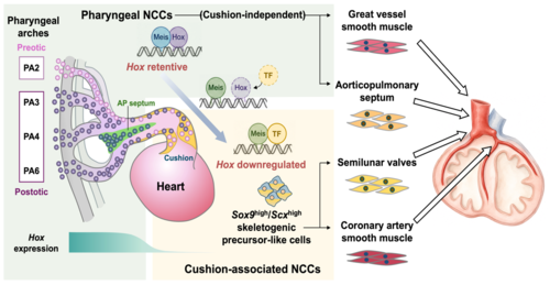
How neural crest cells remodel their regional identity to generate distinct cardiovascular tissues has remained unresolved. By combining single-cell multiomics, spatial transcriptomics, enhancer analyses, and lineage tracing, this study identifies a Hox-Meis-relayed regulatory transition that governs cardiopharyngeal neural crest diversification.
Highlights
➢A comprehensive spatiotemporal atlas links early pharyngeal neural crest cells to late cardiac neural crest derivatives.
➢Loss of Hox programs is coupled to a Meis partner switch that rewires gene regulatory networks during cardiac cushion differentiation.
➢A Meis2-Sox9-Scx regulatory axis defines an intermediate progenitor giving rise to coronary artery smooth muscle and semilunar valve tissues.
Abstract
Neural crest cells (NCCs) are multipotent migratory cells essential for cardiac development, yet the lineage trajectories and gene regulatory networks (GRNs) underlying their differentiation in the cardiopharyngeal region remain unclear. Here, we integrate single-cell RNA-seq, spatial transcriptomics, and multiomic analyses to construct a comprehensive map of NCC lineages in developing mouse cardiopharyngeal tissues. We identify a transition from Hox-positive pharyngeal NCCs to Hox-negative intracardiac populations associated with the outflow tract cushion, accompanied by a shift in Meis transcription factor binding and GRN architecture. By contrast, NCCs forming the aorticopulmonary septum and great vessel smooth muscle retain distinct Hox-codes. A Meis2-Sox9-Scx GRN defines a skeletogenic progenitor-like intermediate state that gives rise to coronary artery smooth muscle and semilunar valves. Our findings suggest that the loss of Hox-dependent regional identity enables pharyngeal NCCs to acquire new fates upon entering the cardiac cushion, providing insight into the developmental origins of coronary and valvular calcification.
Graphical Abstract