
- HOME
- News & Events
- Publications
- 【Publications】Defining compartmentalized stem cell populations with distinct cell division dynamics ...
Publications
【Publications】Defining compartmentalized stem cell populations with distinct cell division dynamics in the ocular surface epithelium
December 18 2020
Aiko Sada
Paper information
Ryutaro Ishii, Hiromi Yanagisawa*, Aiko Sada* (*Corresponding authors)
Defining compartmentalized stem cell populations with distinct cell division dynamics in the ocular surface epithelium
Development. 2020 December 16. doi: 10.1242/dev.197590. URL: https://dev.biologists.org/content/147/24/dev197590.long
Highlights
- This study provides genetic tools to precisely mark and examine the dynamic behavior of heterogeneous stem cell populations in the ocular surface epithelium.
Abstract
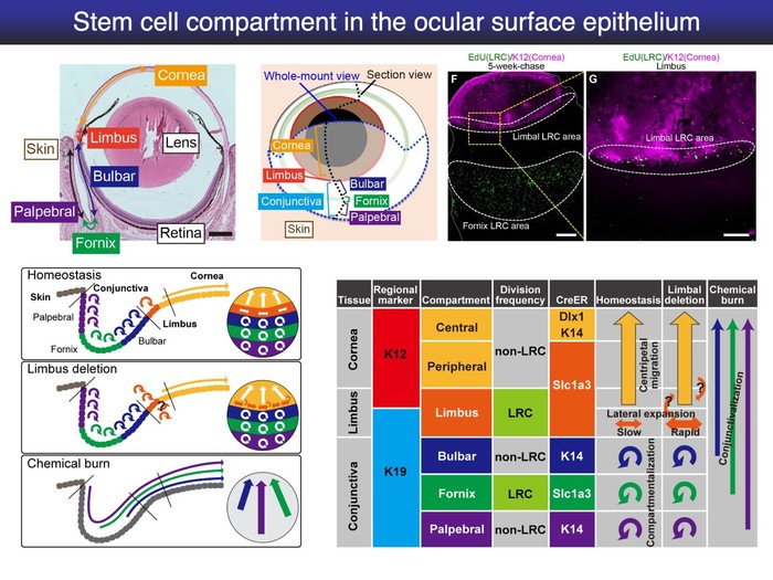
Adult tissues contain label-retaining cells (LRCs), which are relatively slow-cycling and considered to represent a property of tissue stem cells (SCs). In the ocular surface epithelium, LRCs are present in the limbus and conjunctival fornix; however, the character of these LRCs remains unclear, owing to lack of appropriate molecular markers. Using three CreER transgenic mouse lines, we demonstrate that the ocular surface epithelium accommodates spatially distinct populations with different cell division dynamics. In the limbus, long-lived Slc1a3CreER-labeled SCs either migrate centripetally toward the central cornea or slowly expand their clones laterally within the limbal region. In the central cornea, non-LRCs labeled with Dlx1CreER and K14CreER behave as short-lived progenitor cells. The conjunctival epithelium in the bulbar, fornix and palpebral compartment is regenerated by regionally unique SC populations. Severe damage to the cornea leads to the cancellation of SC compartments and conjunctivalization, whereas milder limbal injury induces a rapid increase of laterally expanding clones in the limbus. Taken together, our work defines compartmentalized multiple SC/progenitor populations of the mouse eye in homeostasis and their behavioral changes in response to injury.
![]()