
- HOME
- News & Events
- Publications
- 【Publications】Identification of Stem Cells in the Epithelium of the Stomach Corpus and Antrum of Mic...
Publications
【Publications】Identification of Stem Cells in the Epithelium of the Stomach Corpus and Antrum of Mice
January 5 2017
Motomi Osato
Paper Information
Junichi Matsuo, Shunichi Kimura, Akihiro Yamamura, Cai Ping Koh, Md Zakir Hossain, Dede Liana Heng, Kazuyoshi Kohu, Dominic Chih-Cheng Voon, Hiroshi Hiai, Michiaki Unno, Jimmy Bok Yan So, Feng Zhu, Supriya Srivastava, Teh Meng, Khay Guan Yeoh, Motomi Osato*, Yoshiaki Ito* (*corresponding authors)
Identification of Stem Cells in the Epithelium of the Stomach Corpus and Antrum of Mice
Gastroenterology.152(1):218-231, 2017
Highlights
*An enhancer for Runx1, eR1, marks stomach stem cells.
*eR1-driven oncogene expression promotes cancer development.
Abstract
BACKGROUND & AIMS: Little is known about the mechanisms of gastric carcinogenesis, partly because it has been a challenge to identify characterize gastric stem cells. Runx genes regulate development and their products are transcription factors associated with cancer development. A Runx1 enhancer element, eR1, is a marker of hematopoietic stem cells. We studied expression from eR1 in the stomach and the roles of gastric stem cells in gastric carcinogenesis in transgenic mice.
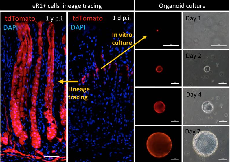
METHODS: We used in situ hybridization and immunofluorescence analyses to study expression of Runx1 in gastric tissues from C57BL/6 (control) mice. We then created mice that expressed enhanced green fluorescent protein (EGFP) or CreERT2 under the control of eR1 (eR1-CreERT2;Rosa-Lox-Stop-Lox [LSL]-tdTomato, eR1-CreERT2;Rosa-LSL-EYFP mice). Gastric tissues were collected and lineage-tracing experiments were performed. Gastric organoids were cultured from eR1-CreERT2(5-2);Rosa-LSL-tdTomato mice and immunofluorescence analyses were performed. We investigated the effects of expressing oncogenic mutations in stem cells under control of eR1 using eR1-CreERT2;LSL-KrasG12D/+ mice; gastric tissues were collected and analyzed by histology and immunofluorescence.
RESULTS: Most proliferation occurred in the isthmus; 86% of proliferating cells were RUNX1-positive and 76% were MUC5AC-positive. In eR1-EGFP mice, EGFP signals were detected mainly in the upper part of the gastric unit, and 83% of EGFP-positive cells were located in the isthmus/pit region. We found that eR1 marked undifferentiated stem cells in the isthmus and a smaller number of terminally differentiated chief cells at the base. eR1 also marked cells in the pyloric gland in the antrum. Lineage-tracing experiments demonstrated that stem cells in the isthmus and antrum continuously gave rise to mature cells to maintain the gastric unit. eR1-positive cells in the isthmus and pyloric gland generated organoid cultures in vitro. In eR1-CreERT2;LSL-Kras G12D/+ mice, MUC5AC-positive cells rapidly differentiated from stem cells in the isthmus, resulting in distinct metaplastic lesions similar to that observed in human gastric atrophy.
CONCLUSIONS: Using lineage-tracing experiments in mice, we found that a Runx1 enhancer element, eR1, promotes its expression in the isthmus stem cells of stomach corpus as well as pyloric gland in the antrum. We were able to use eR1 to express oncogenic mutations in gastric stem cells, proving a new model for studies of gastric carcinogenesis.
![]()