
- HOME
- News & Events
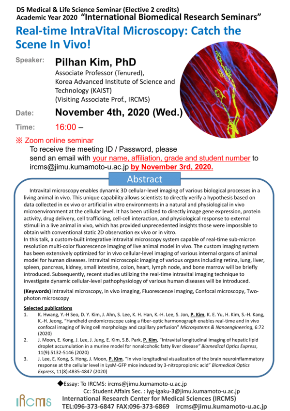
- [Nov.4] D5 Medical & Life Science Seminar-Dr. Pilhan Kim
News & Events
[Nov.4] D5 Medical & Life Science Seminar-Dr. Pilhan Kim
October 16 2020
The "D5 Medical & Life Science Seminar" course will be offered by International Research Center for Medical Sciences (IRCMS). It will run from April 2020 to March 2021, with lectures given by scientists who are affiliated with IRCMS or in collaboration with researchers at IRCMS. The lectures will be given once a month, in English, and by leading scientists in the relevant research field. Students will be taught: 1) how normal physiological functions are maintained in the human body; 2) how these systems become abnormal under certain pathophysiologic conditions; 3) why stem cells are important in animal development and homeostasis; 4) how stem cell-based approaches can help us understand disease mechanisms and find potential cure for diseases related to stem cell malfunction (e.g., cancer, aging).
Date : November 4, 2020 (Wednesday)
Time : 16:00 -
* Zoom online seminar
To receive the meeting ID / Password, please send an email to
ircms@jimu.kumamoto-u.ac.jp by November 3, 2020.
Please include your name, affiliation, grade and student number in your email.
Speaker : Pilhan Kim, PhD
Associate Professor (Tenured),
Graduate School of Medical Science and Engineering (GSMSE)
Korea Advanced Institute of Science and Technology (KAIST)
(Visiting Associate Prof., IRCMS)
Title : Real-time IntraVital Microscopy: Catch the Scene In Vivo!
Abstract :
Intravital microscopy enables dynamic 3D cellular-level imaging of various biological processes in a living animal in vivo. This unique capability allows scientists to directly verify a hypothesis based on data collected in ex vivo or artificial in vitro environments in a natural and physiological in vivo microenvironment at the cellular level. It has been utilized to directly image gene expression, protein activity, drug delivery, cell trafficking, cell-cell interaction, and physiological response to external stimuli in a live animal in vivo, which has provided unprecedented insights those were impossible to obtain with conventional static 2D observation ex vivo or in vitro.
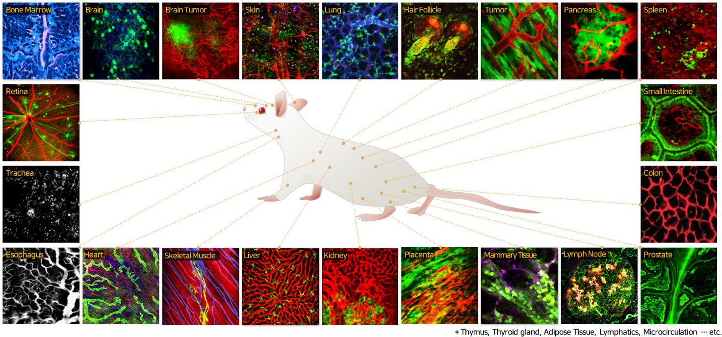
In this talk, a custom-built integrative intravital microscopy system capable of real-time sub-micron resolution multi-color fluorescence imaging of live animal model in vivo. The custom imaging system has been extensively optimized for in vivo cellular-level imaging of various internal organs of animal model for human diseases. Intravital microscopic imaging of various organs including retina, lung, liver, spleen, pancreas, kidney, small intestine, colon, heart, lymph node, and bone marrow will be briefly introduced. Subsequently, recent studies utilizing the real-time intravital imaging technique to investigate dynamic cellular-level pathophysiology of various human diseases will be introduced.
Keyword: Intravital microscopy, In vivo imaging, Fluorescence imaging, Confocal microscopy, Two-photon microscopy
Selected publications
- K. Hwang, Y.-H Seo, D. Y. Kim, J. Ahn, S. Lee, K. H. Han, K.-H. Lee, S. Jon, P. Kim, K. E. Yu, H. Kim, S.-H. Kang, K.-H. Jeong, "Handheld endomicroscope using a fiber-optic harmonograph enables real-time and in vivo confocal imaging of living cell morphology and capillary perfusion" Microsystems & Nanoengineering, 6:72 (2020)
- J. Moon, E. Kong, J. Lee, J. Jung, E. Kim, S.B. Park, P. Kim, "Intravital longitudinal imaging of hepatic lipid droplet accumulation in a murine model for nonalcoholic fatty liver disease" Biomedical Optics Express, 11(9):5132-5146 (2020)
- J. Lee, E. Kong, S. Hong, J. Moon, P. Kim, "In vivo longitudinal visualization of the brain neuroinflammatory response at the cellular level in LysM-GFP mice induced by 3-nitropropionic acid" Biomedical Optics Express, 11(8):4835-4847 (2020)
Flyer:
![]()